Created: May 2007
Note: This article is reproduced here in its original form as written in May 2007. The term Pre-Laminitic Metabolic Syndrome has been superseded by Equine Metabolic Syndrome since this article was written. For a more up-to-date and detailed coverage of this subject, please see my book: ‘Laminitis, a Horse-Centred Approach.’
Introduction
This article assumes the reader is familiar with the mainstream understanding of laminitis. If not, there are plenty of articles on the web that describe what laminitis is.
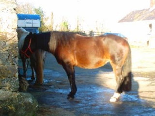
I originally got involved in equine feet because of my mare, Esmé, who has proved to be an incredible teacher when it comes to hoofcare. I struggled for over 4 years to work out why she was never sound – initially in shoes and then (when the shoes contributed to catastrophic hoof pathologies) without shoes. I could make the hooves look better, I could keep her comfortable and even at times fully sound, but it seemed that no matter what I did, any improvements were patchy and limited.
One of the biggest problems I faced was flaring. I would gradually trim the flaring out, but each time I thought I had the flaring under control, the feet would flare dramatically again – sometimes it seemed almost overnight. My initial assumption was that this was somehow to do with moisture levels. Her hoof walls were poor in quality with a lot of cosmetic cracks to let moisture in. Eventually, in desperation, I decided to check back through my records (by then I had 3 years worth of regular photos and notes) to see if they confirmed by suspicions. What I expected to find was that the hooves flared over the winter when she was turned out in wet pasture and that the flare would decrease over the summer when the weather was drier. What I actually found was subtly different.
It seemed that the hooves were always at their best in February /March (when I expected them to be at their worst after a wet winter). The flaring seemed to appear then within a few weeks so that the hooves were at their worst by April/May. They then improved a bit over the summer before going wrong again in the autumn. From October onwards I then saw a steady improvement in flaring, only for things to go wrong again in February the next year. By that time I was working as professional EP and had a number of laminitis cases on my books. I quickly recognised the pattern as matching almost exactly the periods in the year when I saw the most problems with laminitis.
I then started looking at my clients’ horses to see if I was seeing the same pattern and, sure enough, there it was staring me in the face. Not only that, but the horses that matched this pattern were precisely the cases that I most struggled to help. I started trying these horses (including Esmé) on the kinds of dietary changes I normally use for laminitis cases and fairly rapidly started to see impressive results. At the same time I was learning more about diet and how it affects laminitis. Being able to indentify milder symptoms caused/exacerbated by the same dietary problems but at much lower levels allowed me to develop better diets for laminitics of all kinds.
I’ve spent a lot of time looking for references to a condition of this kind in the published literature and have found very little. There is some mention of ‘sub-clinical laminitis’ in the literature, but the very term ‘sub-clinical’ means without visible signs and I was very definitely seeing visible signs (see below for details). It is clear that researchers generally accept that laminitis doesn’t always have to be catastrophic, but there seems very little understanding of exactly what happens in such a case.
Over the last year, I have been studying this condition in my own horses and my clients’. I have been able to build up a picture of a pretty consistent set of clinical signs that are clearly diet related and respond well to the same dietary interventions that work in full blown laminitis. My colleagues and I are now calling this condition Low Grade Laminitis (LGL). We may need to change the name as we discover more about it. I would be extremely interested to talk to any other researchers who are identifying similar patterns.
So what is Low Grade Laminitis?
At a simplistic level, Low Grade Laminitis is just low grade inflammation of the laminar corium. However, unlike the condition that we’ve known and dreaded for generations, in low grade laminitis you don’t get the catastrophic failure of the laminae and the resulting extreme pain and damage to the hoof. There are still a lot of unanswered questions, but the most likely explanation is that a horse with Pre-Laminitic Metabolic Syndrome (PLMS) enters the developmental phase of laminitis but does not progress to the acute phase. From my observations of such cases, I think that many of these horses may have regular bouts of mild inflammation of the laminar corium that come and go through the year. These bouts may last as little as a few hours or as much as a few weeks. Given what we already know about PLMS, the most likely trigger for these bouts is dietary – i.e. eating food that is too sugary for a horse with a compromised sugar metabolism.
This process actually makes a lot of sense in the context of the existing knowledge of and theories about laminitis. There is nothing in the disease models that we currently have that says that the developmental phase of laminitis must automatically lead to the acute phase. If a PLMS horse is already ‘on the edge’ because of a unsuitable diet, it would appear that dietary challenges (maybe something as inocuous as an extra carrot as a treat) can push the horse into a danger zone where the laminar corium becomes inflammed. If those dietary challenges then go away (say the horse doesn’t get a carrot the next day) then the horse can step back from the brink before a catastrophic chain of events starts.
Needless to say, if this theory is correct, a horse exibiting this condition is probably at a much increased risk of developing full blown laminitis and indeed this does seem to be the case. My own horse Esmé has had two mild bouts of full laminitis and I have seen similar things in other horses. That said, there does seem to be quite a large amount of leeway between developing LGL (particularly the milder forms) and developing full blown laminitis. This may suggest that the current assumption that the development phase is short lived and moves almost innevitably towards the acute stage is incorrect. It is possible that this view of the condition has resulted because most laboratory research into the condition is done on animals that are deliberately pushed into laminitis by some major insult (such as being deliberately poisoned or fed excessive amounts of sugary feed).
What happens to the foot in LGL?
Again, there is still much work to do to characterise what really happens in LGL, but here’s my current understanding of what goes wrong as a result of the inflammation. Bear in mind that this represents my current theories and may change as I and other researchers learn more.
Tender footedness
Not every LGL case shows signs of lameness, but many are at least footsore at some stage. Shod horses are far less likely to show any discomfort as shoeing seems to effectively mask a good degree of foot pain. If the laminar corium is inflammed, it is not surprising that the horse can be footsore. We all know how even minor inflammation in our own bodies leads to mild discomfort. It is important however not to confuse tender-footedness from LGL with tender footedness caused by the hoof capsule being in poor shape. Clearly if the sole is very thin (e.g. due to over-trimming), the horse will not be comfortable even with no inflammation present. For this reason, for tender-footedness to be considered an indication of LGL, it needs to be significantly more than would be expected given the externally visible quality of the hoof capsule. In terms of equine podiatry and barefoot horses, an example would be a horse that had a usability score of 6 out of 10 but was still unhappy walking on smooth tarmac.
Where a horse is actively having a LGL attack, it almost always results in an increase in tender footedness in my experience. However, all things are relative – so a horse that is normally fully sound at the gallop on stony tracks might still seem very sound on smooth tarmac despite a mild LGL attack, but might be seen to be a little more hesitant than normal on the stony tracks. In contrast a horse that is only just coping on tarmac without the LGL will appear obviously lame once an LGL attack starts.
Because LGL does minor structural damage to the foot, the tenderness often continues for some time after the LGL attack stops. A mild attack on a foot with a strong hoof may result in discomfort lasting for a week or so even if the attack is stopped (i.e. the diet is changed) within 24-48 hours. As such, tender-footedness is not necessarily an indication that the attack is still current.
Raised Pulses
If you’ve ever hit your thumb with a hammer, you’ll know that it tends to throb – this throbing sensation is caused by increased blood flow to the inflammed tissue. The same happens when the laminar corium is inflammed, but the hoof capsule prevents you feeling the throbbing (the horse feels it though). You can however feel this as an increased strength of pulse in the main arteries to the foot. In traditional laminitis, the digital pulses (best found either side of the pastern just under the fetlock joint) are typically described as ‘bounding’. Vets tend to assume that any pulse that is feelable but not significantly raised is ‘normal’. What I’m finding by studying LGL is that in horses that don’t exhibit this condition, the pulses are difficult to find when the horse is at rest (exercise brings the pulses up a little). In contrast the horses exhibiting active LGL have pulses that are easily found but not necessarily raised to the level that would traditionally be seen as a warning of laminitis or other inflammatory foot condition. Given that the pulses are slightly raised at the point where the corium is actually inflammed, this makes pulse taking an extremely useful technique in picking up LGL early on before too much damage is done.
Bear in mind that other conditions (e.g. abscessing) can also raise the digital pulses. Normally if the pulses are raised in all four feet, this is strongly suggestive of laminitis (the exception would be bruising to all four feet from overuse) whereas a raised pulse in a single foot is far more likely to be an abscess.
I’m seeing some evidence that even after the underlying laminitis attack is brought under control, raised pulses may continue for a while as the damage to the foot heals. Obviously the more severe the damage, the longer it will take for the inflammation to go down after it’s cause is removed and hence the longer the pulses will be raised.
Strong heel first landing and shortened stride
Laminitis tends to make the toes more sore than the rest of the foot, and LGL seems to be no exception to this. One of the first signs of an LGL attack is that the horse starts to put it’s feet down slightly more heel first than normal. A healthy horse should (assuming the horse is walking/trotting with impulsion) put the foot down almost flat but very slightly heel first. The effect is difficult to spot and to the untrained eye often looks like a flat landing. In LGL, the landing becomes more obviously heel first.
Foot pain also typically results in a shortening of the stride length. Trot tends to become flattened (i.e. easier to sit to). In addition, canter departs seem to become more difficult for some horses with canter becoming ‘balled up’ and bucking on transitions more common.
These gait changes are often the first indication that something is going wrong. If you know your horse and the way it moves well, this can be sufficient warning to change the diet and stop something more serious going wrong.
It is not uncommon in my experience for a ‘lazy’ or ‘nappy’ horse to lose these traits when the diet is changed to an anti-laminitis diet. My suspicion is that very mild LGL causes feet to ache slightly which can make a horse reluctant to move forwards with impulsion. Quite a few of my clients have commented that their horses are far more forward since LGL has been addressed and more than one has complained that they can no longer sit to their horses’ trots!
Altered Stance
Even at stand, a horse’s use of his/her feet will alter if they are inflammed. In severe laminitis, you often see a rocked back stance where the horse attempts to take as much weight as possible off the front feet and also off the toes of the hind feet. In LGL, the alterations to stance are far more subtle. Typically, the hind feet are placed slightly further forwards than normal so that the cannon bones are no longer vertical. This tends to result in uneven growth/wear on the hind feet and quite commonly the hind feet struggle to maintain adequate heel height without shoes.
I also commonly see the front feet placed further backwards than usual (i.e. the cannon bones behind the vertical). I am not entirely sure why this happens but it may be part of the process of moving weight backwards onto the hind heels.
One upshot of this stance is that the horse has to use the hind quarters musculature to achieve it (unlike a normal stance where the stay apparatus locks and the horse needs no muscle tension to keep weight balanced on the hind legs). This constant tension in the hind quarters tends to lead to muscle discomfort in the rump and lumbar spine. Stifle problems also seem to be more common in LGL cases (possibly due to uneven muscle development in the hind legs). This muscle tension may well contribute to the alterations in gait described above – especially the reluctance during canter transitions.
Unusual growth rate
Many (but not all) LGL cases show a reduction in growth rate of the hoof. A tiny minority show excessive growth rate. As yet I don’t know what the significance of this is. As such, it is not diagnostic on its own, but as part of the general picture contributes to a suspicion of LGL.
Typically the altered stance described above leads to more stimulus being applied to the heels, especially on the hind feet. This may result in higher growth rate at the heel than at the toe. However, it also typically results in more wear so that the overall effect (especially on the hinds) may be low heels. This difference in heel/toe growth rate, if present, will result in growth rings that are further apart at the heel than at the toe although the effect is usually small enough that it isn’t immediately obvious.
Flaring
There is much debate about the precise mechanisms by which the hoof wall is produced and how it slides past the pedal bone. I don’t want to get into that debate here, however there is one aspect of these mechanisms that is important in LGL. By studying the mechanisms with which the hoof responds to an abscess, you can demonstrate that the hoof wall has a ‘repair mechanism’. Whenever the hoof wall becomes damaged, the laminar corium starts to produce extra horn (which I call ‘repair horn’) that repairs the damage to the wall. When an abscess forms at the sole and blows at the coronary band (the most common type of abscess if the vet hasn’t intervened and drained the abscess at the sole), this repair horn is used to glue the wall back onto the laminar corium and make the hoof strong again. Similarly, where there is a traumatic injury and a section of hoof wall is broken off, the laminar corium will ‘patch’ the hole by producing repair horn. My suspicion is that the trigger for the production of this repair horn is inflammation in the laminar corium. If that is the case, then when the corium is inflammed as a result of LGL, we’d expect to see the corium produce repair horn inside the wall that is distributed according to how much inflammation there is. While I still have work to do to demonstrate that this does indeed happen, what I observe in real cases fits with this theory perfectly.
What I typically see in the days/weeks following a LGL attack is that the wall flares. This flaring alters the angle of the hoof wall from top to bottom simultaneously. Given that it takes 6-7 months for a whole hoof wall to grow down (more in a shod horse), this is too fast to be coming from the coronary band. The only explanation I can find is that the wall is being pushed out from inside by extra repair horn produced at the laminar corium. Once the active LGL stops, the flaring remains but grows down the hoof with the wall. Some months later I find a wall where the top portion is upright and parallel to the pedal bone, but the bottom part is flared. Where the LGL stopped suddenly (maybe because the diet changed for the better), there will be a hard line change of angle in the hoof. It is often far easier to spot LGL once the underlying problem has gone away due to these sudden changes in angle. Spotting it as it first happens takes a good eye for what represents a good shaped hoof (although a front hoof that is significantly longer than it is wide is a good clue). Regular measurements of key parts of the hoof (e.g. ratio of frog length to hoof length) can also help to pick up this flaring early as can taking regular photographs. LGL bouts are often seasonal (being most common in Spring and Autumn) which makes identifying the flaring between bouts easier.
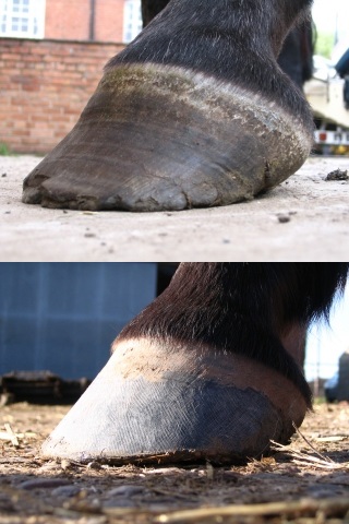
For some reason, some breeds seem to flare more for a given severity of LGL (guaged using comfort levels). In particular, thoroughbreds seem to flare a lot whereas iberian breeds tend to flare less.
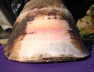
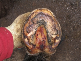
Ring shaped wall bruising
Whenever the laminar corium is inflammed there seems to be a greatly increased risk of bruising in the coronary corium (the tissue that produces the hoof wall – sometimes known as the coronary band). I don’t as yet know whether this damage to the coronary corium is as a result of the same mechanisms that cause the inflammation to the laminar corium or whether this is some kind of mechanical secondary damage due to loss of structure in the laminar corium. Either way, it provides some useful clues, if only in white feet (although in the most extreme cases it is sometimes just possible to see bruising even in a black foot). Unfortunately, the bruise is initially formed behind the periople (the equivalent of a human cuticle that is sometimes confused with the coronary band). As more wall is produced, the bruised horn travels down the wall, taking around 3-4 weeks to appear below the periople – so this sign again only tells us of past LGL bouts. If you have some idea of the growth rate of the hoof, you can estimate how long ago each LGL bout happened by measuring the distance of the bruising from the hairline. This can be useful in trying to determine what is causing the problems.
LGL cases often show bands of orange colouration – sometimes, but not always, with patches of bruising with the band. I now think that this orange colouration represents tissue fluid being released into the horn at the coronary band (similar to a mild grazed knee that weeps tissue fluid but doesn’t actually bleed). With the coronary corium inflammed enough to leek tissue fluid, any further stress on the hoof wall will lead to bruising in that area. This may explain why shod horses with LGL typically have the strongest bruising (and also the most flaring) directly above the nail holes.

White Line Disease
White line disease is an infection of the wall that causes the wall to delaminate. This infection feeds on damaged horn. For the infection to track further than the very surface of the hoof you need either mechanical damage (e.g. from an over-long hoof wall breaking away) or microscopic damage to the hoof wall structure. When the coronary corium gets even very slightly inflammed, blood products (serum and/or red blood cells for example) get incorporated into the horn that the corium produces (giving us the visible bruising described above). As this horn grows down and reaches the bottom of the hoof, it provides the perfect substrate for white line disease to feed on. As I have worked with more and more LGL cases, I’ve noticed that the cases that exhibit white line disease that is difficult to treat almost always have signs of LGL as well. Even more conclusive evidence is that once I have the LGL under control, the white line disease suddenly goes away around about the same time the last bruising and flaring grows off the bottom of the wall.
Sole Bruising
More recently, I’ve been able to demonstrate in cadaver feet that not only do the laminar and coronary coria become inflamed, but the solar corium (which produces the sole) does too. As with the wall bruising, I don’t yet know for sure whether this is a direct effect of the laminitis or a secondary effect due to the solar corium getting overstressed as the rest of the foot fails. However the pattern of bruising is almost always even across the whole sole, suggesting that it is a systemic effect rather than a mechanical one. This solar bruising may well explain the tender-footedness that LGL cases show.
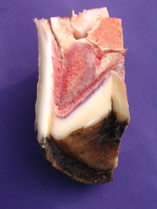
It is rare (although not unheard of) to see this bruising appearing at the surface of the sole. This may be because such bruising provides the perfect feeding ground for infection which will convert the blood products into black slime rapidly and effectively.
Frog Bruising
We’ve already seen bruising in all the other coria that take any kind of mechanical strain while holding up the weight of the horse. It doesn’t seem too much of a leap of faith to assume that the frog also gets bruised. Until recently I’ve struggled to show this (partly due to a shortage of white frogs in my so far limited supply of cadavers). However, a recent live case showed me exactly what I was looking for around 2-3 months after the last bout of laminitis.
Thrush
As with the white line disease, any bruising damage in the frog is likely to be the perfect breeding ground for thrush infections. As you’d expect, LGL cases typically have rampant thrush that can be managed with disinfectants but keeps coming back. After a couple of months of an anti-laminitis diet, all the damaged horn has grown out and the thrush miraculously goes away. LGL cases often have thrush that tracks through the frog in layers, allowing whole sheets of frog (sometimes quite thick) to come away. My suspicion is that this represents a layer of bruising in the frog that gets close enough to the surface for infection to track into it.
Stretched white line
Given that LGL represents inflammation of the laminar corium that doesn’t manage to cause the laminae to actually fail, it makes sense that the worst cases may show a small degree of stretching of the laminae without actually seeing them fail as in full blown laminitis. The insensitive laminae (one half of the laminae) forms half of the white line as it merges with the sole, so a stretched laminae will result in a stretched white line. The worst LGL cases often show a white line that is slightly stretched – especially at the toe area (exactly where laminar wedge forms in full laminitis). If X-rays show clear signs of rotation or sink on the pedal bone, then the diagnosis is clearly of full blown laminitis. LGL cases don’t show these changes on X-ray.
The stretched white line may also be a route for infection to get in and it is common for LGL cases to show significant white line disease actually in the white line (as opposed to in the wall as described above).
Occasionally I’ve seen blood in the white line in these cases (presumably this has come from bruising of the laminar corium some weeks earlier) however this isn’t as common as in full blown laminitis cases.
Abscessing
The inflammation in the various coria forms the perfect home for an abscess to brew. All that is needed is a route for infection to get into the foot. The stretched white line provides just that. As a result, abscesses are more common than normal in LGL cases – especially the more severe ones.
Under-run heels
More recently, I’ve noticed that under-run heels are more common in LGL cases (although this seems also to be related to breed with thoroughbreds being far more prone). The most likely explanation is that the flaring described above contributes to distortion of the heels. However, I have wondered whether the same inflammation processes may directly damage the lateral cartilages in some way.
Flat footedness
The increase flaring tends to pull the white line outwards and in turn the edge of the sole is also pulled outwards. This tends to reduce the amount of concavity. Inflammation of the sole will also result in a thickening of the sole (false sole), especially towards the centre of the sole. If there are under-run heels, these will also contribute to a flattening of the foot’s internal arch. The result is that flat-footedness is more common in LGL cases – especially in breeds that tend to flare a lot with LGL such as thoroughbreds.
Insulin Resistance
The assumption here is that LGL is just a milder form of pasture laminitis. Given that the latest research is suggesting that the underlying condition that predisposes horses to laminitis (PLMS) includes an element of Insulin Resistance (IR), you would expect to see signs of IR in LGL cases. In my experience, not every LGL case shows clear signs of IR, but the majority do.
Conclusions
I’m now convinced that many of the hoof pathologies we commonly see are often as a result of or at least contributed to by LGL. The typical horse that does not do well out of shoes may well be suffering from LGL. In my own practice, I’m increasingly finding that the vast majority of the cases that I struggled with previously now do well without shoes once a suitable diet has been found. Often the improvements gained surprise the owner and sometimes they even surprise me.
As yet we have little hard data on how prevalent this condition is in the UK. 70% of my cases show some signs of LGL – but given the remedial bias to my practice this is not particularly informative. Estimates from my colleagues range from 10% to 50% of horses in the UK. My own suspicion is that the higher figure is probably nearer the mark, with pleasure horses (as opposed to those in heavy work) being the most prone.
It is still early days, but a better understanding of this condition and how to deal with it promises to very significantly improve the health of our horses’ feet.